AI image analysis software is transforming the field of radiology and healthcare. It can detect abnormalities in medical images with high accuracy, reducing the workload of radiologists and improving patient care.
This technology has been shown to detect breast cancer from mammography images with a high degree of accuracy, often surpassing human radiologists.
AI-powered image analysis can also help identify cardiovascular diseases, such as cardiac arrhythmias, from echocardiogram images.
You might like: Genai Image
Radiology and Healthcare
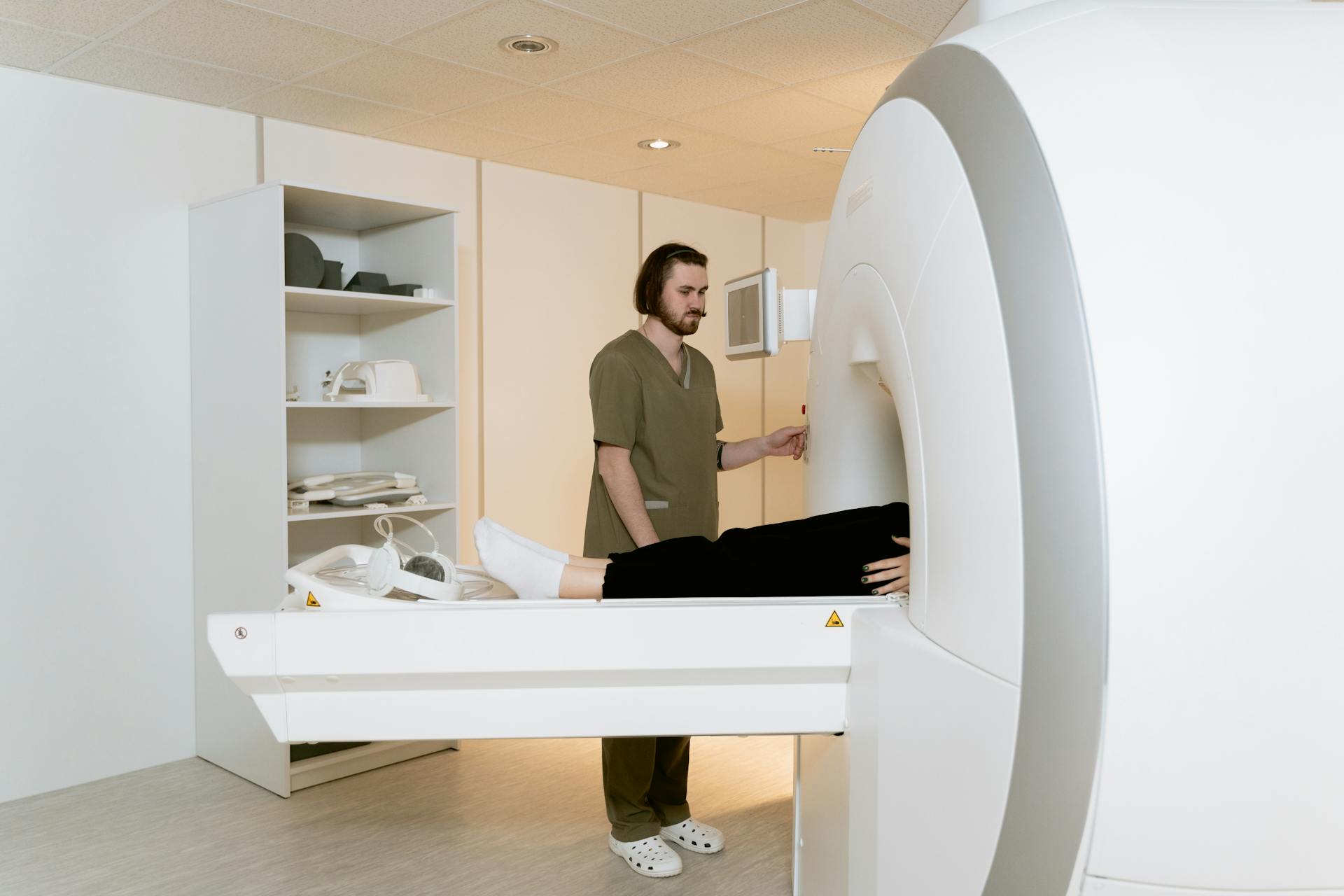
The integration of AI in radiology is transforming the way healthcare providers analyze medical images, leading to more accurate diagnoses and streamlined workflows. This is crucial for improving diagnostics and patient care.
At the heart of this transformation is the development of AI software algorithms that can augment radiologists' expertise and enhance their diagnostic capabilities. The medical imaging AI platform 'Satori' is a prime example of this, offering a comprehensive suite of radiology AI software algorithms.
Accuracy Amplified: AI software can ensure every diagnosis is both detailed and dependable by incorporating its findings with a radiologist's expertise. This is achieved through the integration of AI software with a radiologist's expertise, ensuring that every diagnosis is both detailed and dependable.
Additional reading: Ai Medical Dictation Software
Patient-Centric Care: AI-enhanced radiology software can facilitate timely interventions by enabling early disease identification, setting new standards in patient care. This is made possible by the array of tools offered by Satori, which enable healthcare setups to capitalize on early disease identification.
The benefits of AI in radiology are numerous, and its impact is already being felt in the healthcare industry. From image interpretation to patient positioning, AI is revolutionizing the field of radiology.
On a similar theme: Ai Radiology Software
Getting Started
If you're new to AI image analysis, it's essential to start with a solid foundation. Satori, a Radiology AI software, is a great place to begin. Every algorithm on offer has been crafted with care, rigorously tested, and proven to deliver tangible benefits to radiology practices.
Satori's platform is designed to be user-friendly, making it easy to dive into the world of AI image analysis. You can discover the vast possibilities these algorithms bring to the table and initiate your journey into the AI realm of radiology.
Check this out: Ai Image Training
To get started, you'll need a platform that offers state-of-the-art image visualization and analysis tools. Aivia Go is a great option, providing multiple AI-powered features in a single platform. This includes simple segmentation workflows and batch processing capabilities that yield results faster.
Aivia Go supports a range of applications from 2D to 5D image analysis tasks such as detection and tracking. It's ideal for research laboratories and imaging core facilities, and no computer expertise is required.
Here are some key features of Aivia Go:
- AI-powered classifiers
- 12+ image analysis recipes
- 2D -5D image visualization
For more advanced users, Aivia Apex is a comprehensive microscopy image analysis solution that provides a wide range of image analysis applications. It's ideal for large research groups and core imaging facilities, and its Floating License Manager allows a site to run all licenses on any machine on their local network.
Aivia Apex offers a variety of image analysis recipes, including nuclei count and tracking, cell count and tracking, and particle count and tracking. These recipes can be deployed for the most popular analysis applications, making it a powerful tool for researchers.
Additional reading: Software for Ai Data Analysis
Software Features
HALO offers a wide range of image analysis recipes for various applications, including cell counting, tracking, and proliferation assays, as well as neurite outgrowth and stem cell colony detection.
These recipes can be deployed in both 2D and 3D, making it a versatile tool for researchers. Additionally, HALO allows for the tracking of cells, nuclei, and particles in 2D and 3D.
Some of the specific features of HALO include real-time tuning tools, annotation tools, batch analysis capabilities, and a multi-panel figure maker for easy publication.
Here are some of the key features of HALO:
- Cell counting, tracking, and proliferation assays
- Neurite outgrowth and stem cell colony detection
- Real-time tuning tools
- Annotation tools
- Batch analysis capabilities
- Multi-panel figure maker
These features make HALO a user-friendly and powerful tool for image analysis, with great technical support to boot.
Leverage Integrated Tools
The HALO platform offers a range of integrated AI tools that can be used to analyze images and extract valuable insights. You can leverage pre-trained deep-learning networks for optimized nuclear and membrane segmentation directly in the HALO platform.
Suggestion: Ai Software Platforms
HALO's cell analysis modules in brightfield or fluorescence are particularly useful for applications such as Multiplex IHC and Highplex FL. These modules allow for accurate analysis of immunolabeling and can extract cell features integrating spatial information.
You can also use HALO's real-time tuning tool to optimize analysis parameters and see the effects in real-time. This feature is especially useful for complex image analysis tasks.
Here are some of the key features of HALO's integrated AI tools:
- Pre-trained deep-learning networks for optimized nuclear and membrane segmentation
- Cell analysis modules in brightfield or fluorescence
- Real-time tuning tool for optimizing analysis parameters
- Annotation tools for selecting regions of interest
- Batch analysis for processing multiple images at once
By leveraging these integrated AI tools, you can streamline your image analysis workflow and extract valuable insights from your data.
Compatibility
HALO is compatible with a wide range of image and digital slide formats.
You can import files in non-proprietary formats like JPG and TIF, as well as proprietary formats from leading manufacturers.
HALO supports files from Nikon, 3DHistech, Akoya, Olympus, and many others.
The software is compatible with over 20 different file formats, including MRXS, QPTIFF, and VSI.
Readers also liked: What Software Opens Ai Files
Some of the formats HALO can read include ND2, OME.TIFF, and MRXS.
HALO can also import DICOM files, which is a standard format used in medical imaging.
Here's a list of some of the formats HALO is compatible with:
- Non-proprietary (JPG, TIF, OME.TIFF)
- Nikon (ND2)
- 3DHistech (MRXS)
- Akoya (QPTIFF, component TIFF)
- Olympus / Evident (VSI)
- Hamamatsu (NDPI, NDPIS)
- Aperio (SVS, AFI)
- Zeiss (CZI)
- Leica (SCN, LIF)
- Ventana (BIF)
- Philips (iSyntax, i2Syntax)
- KFBIO (KFB, KFBF)
- DICOM (DCM)
Efficient Workflows
Efficient workflows are crucial for getting the most out of your ai image analysis software. HALO, for example, offers a streamlined workflow for co-registration and cellular analysis of immunofluorescence images from cyclically stained and imaged slides.
HALO's intuitive platform allows analysts at all levels to be up and running with basic analysis functions in just a couple of hours, with no prior image analysis experience required. This is made possible by HALO's purpose-built modules that simplify the analysis workflow and eliminate the need to build algorithms from scratch.
Some of the key features of efficient workflows in ai image analysis software include:
- Batch analysis for increased productivity
- Workflow tools for efficient tissue microarray and serial stain analysis
- Collaborative tools for working with others on image analysis projects
These features can help you and your team work more efficiently and effectively, freeing up time to focus on more important tasks like innovation and discovery.
Efficient Workflows
HALO offers a streamlined workflow for co-registration and cellular analysis of immunofluorescence images from cyclically stained and imaged slides.
The Lunaphore COMET instrument enables a high-throughput workflow for phenotypic and spatial analysis of cells in the tumor microenvironment, when paired with HALO and HALO AI.
You can analyze cellular interactions and measure the distances between different compartments down to the channel level with Elevate for CellBio.
Aivia's AI-powered analysis capabilities leverage a biologist's expertise to generate robust and reproducible segmentation results.
Aivia's powerful and fast 2-5D visualization and analysis unlocks all the value of your data - within a single platform.
Here are some key features of efficient workflows:
- Multi-core processing and batch analysis
- Workflow tools for efficient tissue microarray and serial stain analysis
- Co-registration and cellular analysis of immunofluorescence images
- High-throughput workflow for phenotypic and spatial analysis of cells
- Streamlined workflow for extracting information from hyperplex datasets
Daily Study Coordination
Daily Study Coordination is a breeze with HALO & HALO-Link.
We use HALO & HALO-Link daily to coordinate studies for our analysts and pathologists.
HALO is a whole-slide image analysis platform that is designed to work how pathologists classify & score images using a microscope.
It can be learned in a day to analyze brightfield & fluorescent whole slide images and TMAs.
File sharing through HALO-Link lets users access studies through a simple web-interface sharing a common database.
HALO-Link enables seamless collaboration and streamlines the workflow.
Expand your knowledge: Getty Generative Ai
Customer Testimonials
Our customers rave about the benefits of using ai image analysis software to streamline their workflows. They've seen significant improvements in efficiency and productivity.
Indica Labs' solutions have been a game-changer for many of them, allowing them to focus on more complex tasks and projects.
Customers have reported a notable reduction in the time spent on manual data analysis, freeing up resources for more valuable work.
Reading independent reviews on our HALO image analysis platform is a great way to learn from others who have successfully implemented these solutions.
Research and Development
In the world of research and development, having the right tools can make all the difference. HALO has become a fundamental tool in our research activity, allowing for accurate analysis of both brightfield and fluorescence-based microphotographs or slide scans.
With HALO, you can easily extract cell features integrating spatial information, enabling detailed spatial interaction analyses. This is particularly useful in fields like cancer research, where understanding immune responses is crucial.
Aivia 14 offers a suite of novel tools for exploring and visualizing complex data and spatial relationships in 2D and 3D multiplexed images.
The improved deep-learning model in Aivia 14 accelerates cell detection by up to 78% for 3D objects. This is a significant improvement, allowing researchers to process large datasets much faster.
Aivia 14 also accurately segments cells with different morphologies in 2D and 3D multiplexed images. This is a critical feature for researchers working with complex data.
Here are some key features of Aivia 14:
- Improved deep-learning model accelerates cell detection by up to 78% for 3D objects
- Accurately segment cells with different morphologies in 2D and 3D multiplexed images
- Discover the cell phenotypes in your image using AI-powered phenotyping or data-driven unsupervised automatic phenotyping
- Interactively explore phenotypes and gain a deeper understanding of 2D and 3D multiplexed image data using dendrogram and dimensionality reduction tools
Modules and Tools
HALO offers a range of modules and tools for image analysis, including the Multiplex IHC and Highplex FL modules for optimized nuclear and membrane segmentation.
You can filter by category and view additional options using the arrows at the bottom of the list. Some popular HALO modules include Quantify expression of up to five brightfield stains in any cellular compartment, and Quantify expression of an unlimited number of biomarkers in any cellular compartment.
The HALO platform also includes a TMA Add-on for automated, high-throughput segmentation and batch analysis of whole slide TMA images. This can be a productivity-enhancing workflow for tissue microarray analysis.
Some HALO modules, such as the Plot cells and objects module, allow you to perform nearest neighbor analysis, proximity analysis, and tumor infiltration analysis. Others, like the Simultaneously analyze module, enable you to analyze up to three chromogenic and/or silver-labelled DNA or RNA ISH probes on a cell-by-cell basis.
Here are some of the HALO modules and their features:
Aivia also offers a range of tools and features, including parameter-free image segmentation and easy training and updating of deep learning models. The platform includes 22 applications and 20 pre-trained deep learning models, and supports over 45 microscopy file formats.
Expand your knowledge: Geometric Feature Learning
Frequently Asked Questions
What is the best image analysis software?
There isn't a single "best" image analysis software, as the choice depends on the specific needs of your project. However, popular options include ImageJ/FIJI, Cell Profiler/Cell Analyst, Neuronstudio, and VIAS, each with unique features and applications.
What is the AI that can read a picture?
Image recognition AI, also known as computer vision, is a technology that enables machines to interpret and understand visual content from images and videos
Featured Images: pexels.com


